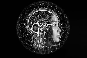
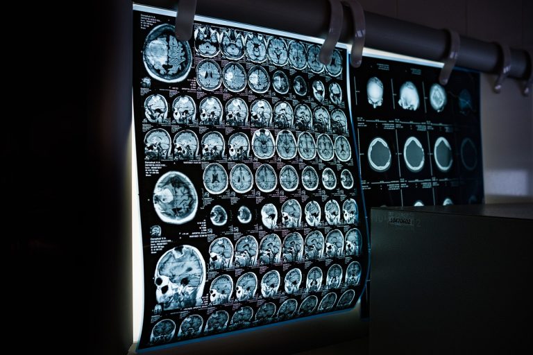
The military and the private sector have both embraced technical breakthroughs in the digital age. One industry has, however, been in the vanguard of swiftly embracing the most recent technological change. More than 580 healthcare technology agreements worth a staggering $83 billion were struck in the US between 2014 and 2018. Three important categories were focused on: new care models, data and analytics, and patient involvement. The improvement of the patient experience and the precision of diagnoses are the main objectives of integrating high-tech solutions in the healthcare sector. We will examine the enormous effects of this technological revolution on the innovative medical imaging industry today.
Importance of Medical Imaging: Enhancing Diagnoses and Transforming Patient Care
According to Talissa AItes, Chair of Radiology and Pediatric Radiologist at MU Healthcare, innovative medical imaging is essential for managing various types of patients, including those with cancer, adults, and orthopedic cases. She emphasizes that without imaging, proper patient management becomes challenging. Innovative medical imaging serves as the foundation for diagnosing numerous diseases, as stated by Bob Kittyle, a Radiologist at Florida Hospital Waterman. He further explains that many diseases are initially detected through innovative medical imaging, and high-quality equipment facilitates early or initial-stage disease diagnosis.
However, to ensure effective results, it is crucial to have qualified radiologists, cooperative patients, and skilled physicians. Having certified physicians with expertise in multiple modalities is essential for obtaining accurate medical imaging results. This enables radiologists to collaborate with technicians to enhance image quality, resulting in early diagnosis. Furthermore, excellent equipment allows radiologists to work closely with technicians to produce exceptional images, thereby facilitating early diagnosis and guiding patient care, including surgical or interventional radiology interventions. Professor Peter Soyer from the University of Queensland Diamantina Institute highlights the challenge of distinguishing between multiple pigmented spots on a patient’s back to identify which spot may have melanoma.
Dr. Shakes Chandra, from the School of Information Technology and Electrical Engineering, has developed innovative deep-learning techniques for medical image analysis. These techniques aim to create new AI methods that analyze, extract, and distill information crucial for healthcare professionals from diagnostic, treatment, and assessment images of various human diseases and injuries. Analyzing medical images requires extensive experience and training. However, AI accelerates this process and enables faster data processing. Moreover, it empowers us to train the data, unlocking new possibilities beyond our previous expectations.
In summary, the integration of AI, Machine Learning, and advanced 3D imaging holds great promise in expediting diagnosis, optimizing resource utilization, and enhancing disease detection capabilities. Professor Soyer and Dr. Chandra’s work exemplify the potential of these technologies to revolutionize medical image analysis, ultimately benefiting healthcare professionals and improving patient outcomes.

Magnetic Resonance Imaging (MRI):
Magnetic Resonance Imaging (MRI) is one of the most fascinating diagnostic procedures known in the medical world and is considered at the forefront of innovative medical imaging technology. The images produced by the large tube can be breathtaking and provide crucial information on many diseases. An MRI machine produces sectional images in any spatial direction. It can be used to visualize strokes, inflammation, cancer, as well as damage to muscles, tendons, and ligaments. Another specialty of MRI is imaging blood vessels and the heart.
Computed tomography (CT):
A CT scan is an X-ray procedure that creates cross-sectional images with the help of computer processing. CT images are more detailed than conventional X-ray images and can reveal bones as well as soft tissues and organs. In contrast to a CT scanner, which spins around the patient while shooting narrow beams of X-rays, a conventional X-ray employs a stationary tube that sends X-rays only in one direction. They are special digital X-ray detectors located directly opposite the X-ray source.
The X-rays are transferred to a computer by the detectors, which catch them as they travel through the patient. Image slices can either be displayed individually in two-dimensional form or stacked together to generate a three-dimensional image that can reveal abnormal structures to help the Physician plan and monitor treatments.

Ultrasound Imaging
A vital part of most ultrasound systems is a transducer, which uses a number of piezoelectric crystals. These crystals have a special characteristic called piezoelectricity, which vibrates whenever an electric input is detected. This extraordinary quality serves as the foundation for ultrasonic technology.
Cutting-Edge Technologies in Medical Imaging: Advancing the Frontiers of Diagnosis and Care
Innovative medical imaging techniques like ultrasound, CT, and PET are wonders of science in detecting diseases before they become serious, but they might not offer the degree of detail required to give us a clear picture at the molecular level. Advanced methods are required to achieve deeper knowledge. With the help of these sophisticated techniques, scientists can gain insightful information that helps them identify prospective drug candidates. Here are their major advancements that hold tremendous promise for improving patient outcomes and reshaping the future of healthcare.
3D Imaging:
The 3D medical imaging technology allows doctors not only to see three-dimensional pictures but to interact with them as if they were real using devices such as virtual reality viewers as well as styluses r other hardware that provides tactile feedback. Doctors can take a tour of a patient’s brain for example and even cut into virtual tissue. The technologies come in a variety of forms from fully immersive virtual-reality studios to simulations that use virtual reality viewers such as the rift headset from Oculas. 3D imaging can reduce surgical planning time by 40% and increase surgical accuracy by 10%.

Artificial Intelligence (AI) in Imaging:
Computer algorithms today are performing incredible tasks with high accuracy, at a massive scale, using human-like intelligence, and this intelligence of computers is often referred to as AI. We still face massive challenges in detecting and diagnosing several life-threatening illnesses such as infectious diseases and cancer. Every year, thousands die due to liver and oral cancer. The best way to help patients is to perform early detection of diseases. Traditional medical imaging calls for a very resource-intensive process, requiring both expert physicians and expensive medical imaging technologies, which is not practical in the developing world and in fact in many industrialized nations as well. Can using Artificial Intelligence solve this problem?
MIT Media Lab Principal Investigator and Principal Research Scientist Practik Shah has developed a technology using an unorthodox AI approach, that requires only as few as 50 images to develop a working algorithm and can even use photos taken on doctors’ cell phones to provide a diagnosis. It means that the algorithms can accept very simple white light photographs from the patient instead of expensive medical imaging technologies.

Molecular Imaging:
Molecular Imaging provides physicians with the incredible opportunity to observe the inner workings of the body with exceptional spatial resolution. It enables them to delve into the biochemical processes that contribute to the onset of diseases, creating a promising landscape that embraces the remarkable achievements of molecular medicine and biology. The utilization of specifically designed drugs, targeted toward the receptors expressed by various diseases in cellular systems, represents the future of diagnostic and therapeutic medicine.
When combined with high-resolution detectors and advanced systems for data transmission, leveraging the power of the internet and mobile devices, we can significantly enhance outcomes, such as minimizing costly hospital stays and reducing treatment times.
The Revolutionary Impact of Innovative Medical Imaging in Healthcare
Over the past ten years, thanks to innovation and the use of artificial intelligence (AI), the medical world has seen major breakthroughs in radiology and imaging. These advancements have significantly enhanced patient outcomes and treatment.
Radiology has enhanced diagnostic and treatment choices by using AI to get more accurate results. AI systems can instantly analyze millions of medical photos, aiding in the early diagnosis of diseases. For example, AI enhances the accuracy of mammograms for the detection of breast cancer and enhances the interpretation of MRIs for the diagnosis of prostate cancer.
Additionally, MRI methods have improved, becoming quicker, safer, and more effective. Faster scans with detailed images are made possible using “compressed sensing” MRI, which is advantageous for youngsters and individuals with mobility concerns. The Whole-Body Magnetic Resonance Imaging (WBMRI) test is incredibly sensitive and can help diagnose and cure diseases like multiple myeloma at an early stage.
Dual-energy CT scans have been created to reduce radiation exposure during CT scans. These scans deliver excellent images while lowering radiation exposure. They aid radiologists in precisely identifying healthy and sick tissues.
Particularly in rural and distant places, teleradiology has been a critical component of the digital transformation of healthcare systems. It reduces waiting times and fills the gap between patients and radiologists, enhancing access to imaging services. Teleradiology facilitates quicker diagnoses, which lessens the workload for medical professionals.
The enormous volumes of data produced by these cutting-edge technologies must now be safely stored, managed, and shared, and cloud-based management solutions are therefore essential. They minimize the chance of data loss or breaches and provide simple access to medical photos from anywhere.
In conclusion, technological and AI-driven advances in radiology and innovative medical imaging have transformed the medical sector. Diagnoses, options for therapy, and patient outcomes have all been enhanced by these developments. Teleradiology, dual-energy CT scans, faster MRI procedures, AI-driven analysis, and cloud-based storage options have all improved access to imaging services and improved healthcare delivery.
Final Thoughts:
Innovative medical imaging technologies have revolutionized healthcare. From MRI, CT, and ultrasound to 3D imaging, AI, and molecular imaging, these advancements have improved diagnoses, treatment planning, and interventions. They have accelerated data processing, enabled early disease detection, and enhanced accessibility through cloud-based storage. Teleradiology has reduced waiting times and improved access, benefiting both patients and medical professionals. These technological breakthroughs have transformed patient care, promising a future of improved outcomes, optimized resource utilization, and reshaped medicine. Revolutionary medical imaging is changing our perspective of how we diagnose, treat, and care for patients worldwide.

